Sail E0 Webinar

11th And 12th > Biology

BODY FLUIDS AND CIRCULATION MCQs

Total Questions : 60 | Page 1 of 6 pages
Question 1.


If coronary circulation is removed from the human body, which of the following effects do you think it will have on the body?


  1.     Nutrients from digestive tract will not be transported to the liver.
  2.     Mixing of deoxygenated and oxygenated blood in the heart.
  3.     The heart muscles will stop contracting.
  4.     No transmission of nerve impulses from vasomotor centre to the sympathetic nerves.
 Discuss Question
Answer: Option C. -> The heart muscles will stop contracting.
:
C

Coronary circulation is responsible for supplying blood to the muscles of the heart. From the aorta coronary arteries supply oxygenated blood to the heart muscles while coronary veins carry deoxygenated blood away from the heart muscles. If this circulation is removed our heart muscles will not receive oxygen and will eventually stop contracting.


Question 2.


Which of the following is not correctly matched?


  1.     Systemic circulation - generates efficient waveforms
  2.     Tachycardia - Heart rate is 70-75 beats
  3.     Bradycardia - Heart rate falls below 60 beats
  4.     Atrial fibrillation - generates inefficient waveforms
 Discuss Question
Answer: Option B. -> Tachycardia - Heart rate is 70-75 beats
:
B

When the heart pumps blood into the systemic circulation, it does so with a huge amount of pressure. This pressure becomes a waveform that travels through the arteries ahead of the actual column of the blood which is pumped out. This waveform creates the throbbing sensation we feel when we palpate peripheral arteries. This is what we call the pulse. The normal pulse or heart rate is around 70 to 75 times per minute averaging at 72 beats per minute. Atrial fibrillation is where the ineffective pumping of the heart becomes insufficient to produce waveforms that can reach the periphery. Any heart rate above 100 beats per minute is considered tachycardia and anything below 60 beats per minute is called bradycardia.


Question 3.


Which of the following is/are the composition of the fluid in lacteals?


  1.     Fats and chyme
  2.     Chyme and chyle
  3.     Chyme and lymph
  4.     Lymph and fat
 Discuss Question
Answer: Option D. -> Lymph and fat
:
D

Fats are absorbed from the digestive system by lymphatic vessels called lacteals, which contain a fluid mixture of fats and lymph called chyle. Fats are not absorbed in the blood capillaries as they are too small. So the fats enter the large pores of lacteals instead.


Question 4.


Veins do not require thick walls


  1.     as they do not have to work harder
  2.     they are required only occasionally
  3.     blood pressure declines while it gets to veins
  4.     blood pressures is immense
 Discuss Question
Answer: Option C. -> blood pressure declines while it gets to veins
:
C

Veins carry blood back into the heart and for it less pressure is required. Therefore, veins have comparatively thinner walls than arteries.


Question 5.


Which of the following statements is incorrect?


  1.     A person of ‘O’ blood group has anti ‘A’ and anti ‘B’ antibodies in his blood plasma
  2.     A person of ‘B’ blood group can’t donate blood to a person of ‘A’ blood group
  3.     Blood group is designated on the basis of the presence of antibodies in the blood plasma
  4.     A person of AB blood group is universal recipient
 Discuss Question
Answer: Option C. -> Blood group is designated on the basis of the presence of antibodies in the blood plasma
:
C
Blood Group is decided by the Antigen that is present on the RBCs.
Question 6.


Which blood vessel wall layer is correctly matched with the description about its tissue structure?


  1.     Tunica externa (adventitia) - consists of smooth muscle tissue
  2.     Tunica interna (intima) - consists of only a thin layer of connective tissue
  3.     Tunica media - consists of simple squamous epithelium
  4.     Tunica media - consists of smooth muscle and elastic connective tissue, the amounts and proportions of each depending on vessel type
 Discuss Question
Answer: Option D. -> Tunica media - consists of smooth muscle and elastic connective tissue, the amounts and proportions of each depending on vessel type
:
D

The tunica media is the middle layer of an artery or vein. It lies between the tunica intima on the inside and the tunica externa on the outside.The tunica media, or middle coat, is made up principally of smooth (involuntary) muscle cells and elastic connective tissues arranged in roughly spiral layers.It has the autonomic control of which can alter the diameter of the vessel and affect the blood pressure.


Question 7.


Match the following Transcellular fluids with their functions:


Match The Following Transcellular Fluids With Their Function...


  1.     (i)a, (ii)b, (iii)c,(iv)d
  2.     (i)d, (ii)c, (iii)b,(iv)a
  3.     (i)c, (ii)d, (iii)a,(iv)b
  4.     (i)b, (ii)a, (iii)d,(iv)c
 Discuss Question
Answer: Option B. -> (i)d, (ii)c, (iii)b,(iv)a
:
B

The synovial fluid acts as a lubricant in the joints of the bones. The intraocular fluid present in the eyes supplies nutrition and also maintains the pressure in the eye.


The cerebrospinal fluid acts primarily as a shock absorber but also supplies nutrition. The intracellular fluid, which is present inside the cell, maintains the shape and size of cell.


Question 8.


The first sound of the heart beat occurs during 1______, associated with the closure of  2_______, and 3______, The second sound of the heart beat occurs during 4_____, associated with the closure of  5______, and 6______.


  1.     1.Isovolumetric contraction   2.Mitral valve   3.Tricuspid valve  4. IsovolumetricRelaxation  5. Aortic valve   6. Pulmonary Valve
  2.     1.Isovolumetric Relaxation  2. Aortic valve  3. Pulmonary Valve  4. Isovolumetric contraction   5.Tricuspid valve   6.Mitral valve
  3.     1.Isovolumetric contraction   2.Mitral valve   3.Aortic valve   4.Isovolumetric Relaxation   5.Tricuspid valve   6.Pulmonary Valve
  4.     1.Isovolumetric Relaxation   2.Mitral valve   3.Aortic valve   4.Isovolumetric contraction   5.Tricuspid valve  6. Pulmonary Valve
 Discuss Question
Answer: Option A. -> 1.Isovolumetric contraction   2.Mitral valve   3.Tricuspid valve  4. IsovolumetricRelaxation  5. Aortic valve   6. Pulmonary Valve
:
A

The most fundamental heart sounds are the first and second sounds, usually abbreviated as S1 and S2. S1 is caused by closure of the mitral and tricuspid valves at the beginning of isovolumetric ventricular contraction S1 is normally slightly split (~0.04 sec) because mitral valve closure precedes tricuspid valve closure; however, this very short time interval cannot normally be heard with the stethoscope and hence only a single sound is perceived. S2 is caused by closure of the aortic and pulmonic valves at the beginning of isovolumetric ventricular relaxation


Question 9.


Which of the following is not true about the heart’s conduction system?


  1.     Other muscles receive impulses through the neurons from the brain, but heart produces its own potential
  2.     The SA node generates action potentials higher than all the other parts of the conducting system.
  3.     The SA node generates specifically 70 action potentials per minute.
  4.     If all the other portions of the conducting system other than the SA node, produce action potentials at a much slower rate, it would maintain our heart rate.
 Discuss Question
Answer: Option D. -> If all the other portions of the conducting system other than the SA node, produce action potentials at a much slower rate, it would maintain our heart rate.
:
D

Although all of the heart's cells have the ability to generate the electrical impulses (or action potentials) that trigger cardiac contraction, the sinoatrial node normally initiates it, simply because it generates impulses slightly faster than the other areas with pacemaker potential, all the other portions of the conducting system other than the SA node, produce action potentials at a much slower rate, it would maintain our heart rate.


Question 10.


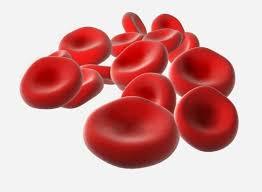
Assertion (A) – RBCs are circular and biconcave in shape lacking all cell organelles.


Reason (R) - Circular and biconcave shape increase the surface area for oxygen binding and the lack of organelles results in no self-utilisation of the oxygen being carried.


  1.     Both A and R are correct and R is the correct explanation of A.
  2.     A is correct but R is wrong
  3.     A is wrong but R is correct
  4.     Both A and R are wrong
 Discuss Question
Answer: Option A. -> Both A and R are correct and R is the correct explanation of A.
:
A

RBCs are usually biconcave and circular in shape. This gives them more surface area resulting in efficient binding of oxygen molecules. Moreover, due to the lack of organelles, the RBCs cannot and need not utilise the oxygen they carry for themselves. This also gives them flexibility.

Assertion (A) – RBCs Are Circular And Biconcave In Shape L...


Latest Videos

Latest Test Papers